2026-06-04
Why CT and X-ray Scans Are Essential Before Facial Contouring Surgery: Checking Facial Nerves and Bone Structure
When planning facial contouring or orthognathic surgery, CT and X-ray scans are the first step toward a safe procedure. In this article, we explore the differences between a 2D X-ray and a 3D CT scan, and explain in detail how identifying the location of the inferior alveolar nerve (facial nerve) impacts surgical outcomes and safety.
Why CT and X-ray Scans Are Necessary Before Facial Contouring Surgery
When consulting for facial contouring or orthognathic surgery (double jaw surgery), one of the very first steps is taking a CT (Computed Tomography) and X-ray (radiograph). This is not just a simple procedure, but a crucial step to establish a safe surgical plan by meticulously analyzing each individual's unique facial bone structure and proportions, as well as the location of major nerve lines.
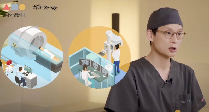
What is the Difference Between an X-ray and a 3D CT?
The reason clinics use both types of equipment is that the nature of the information each provides is different. The two devices have a mutually complementary relationship.
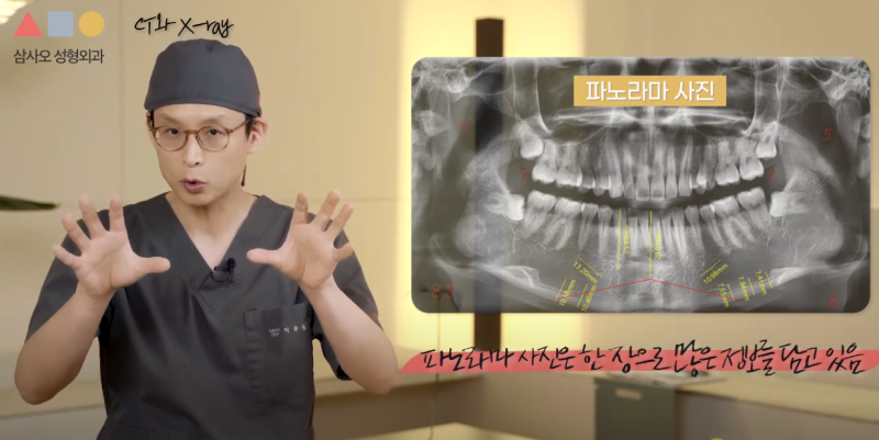
1. X-ray: The Foundation of 2D Planar Data
An X-ray is the most widely used 2D radiograph. Taken from the front or side, it shows the overall contour of the bones on a flat plane.
- Advantages: A panoramic photo that unfolds the entire jawbone is excellent for checking dental alignment, chin distance, and overall asymmetry.
- Characteristics: It reveals not only the bones but also the silhouette of the skin and soft tissues to some extent, serving as a reference for predicting external changes.
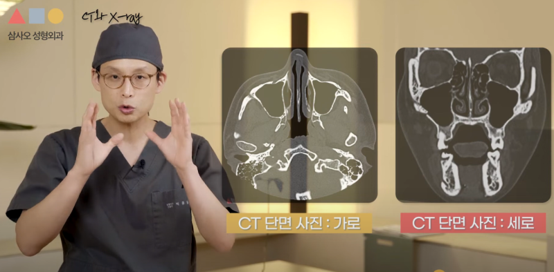
2. 3D CT: Precise Analysis of 3D Volumetric Structure
A 3D CT scan creates a three-dimensional image by reconstructing cross-sectional images taken from multiple angles. Because it renders the patient's facial bones in 3D, closely resembling reality, it allows for an intuitive understanding for both the medical staff and the patient.
- Advantages: It allows for meticulous measurements of the cheekbone width, square jaw thickness, and bone density down to the millimeter (mm).
- Nerve Line Identification: In particular, it can trace the pathway of the inferior alveolar nerve (lower jaw nerve) in cross-sections, playing a crucial role in minimizing the risk of nerve damage during surgery.
Key Information Verified Through Precision Scans
Based on the scan results, a board-certified plastic surgeon comprehensively analyzes the following factors to determine the operable range and surgical design.
- Measuring Nerve Line Height: Determines the safe limit for bone resection during square jaw reduction surgery.
- Skeletal Asymmetry Analysis: Quantifies the differences in size and position of the left and right bones to establish a correction plan.
- Relationship Between Teeth and Skeleton: Assesses the degree of front teeth protrusion and the distance to the lips to enhance the precision of protruding mouth or orthognathic surgery.
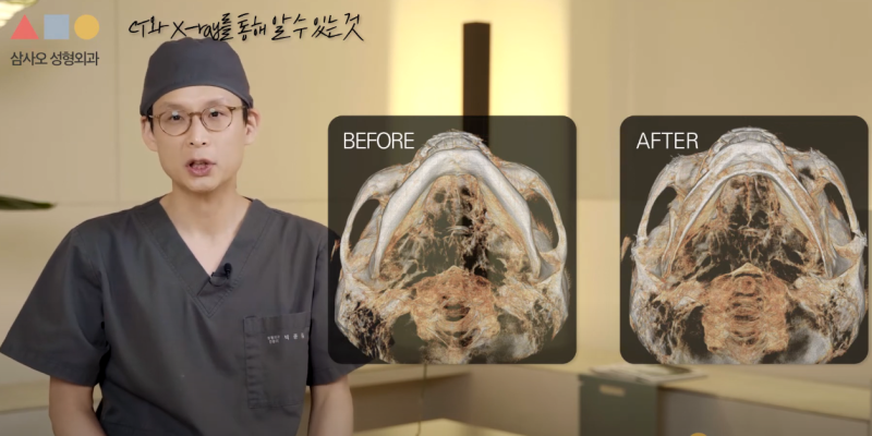
- Pre- and Post-operative Comparison: Allows for a clear comparison of the amount of cheekbone reduction through various angles of the 3D CT (such as the worm's-eye view).
Will Radiation Exposure Affect My Health?
Many people worry about cumulative radiation, but medical CTs and X-rays have their emission levels optimized to the exact amount necessary for an accurate diagnosis. The amount of radiation exposed during a typical plastic surgery examination is very low, even when compared to the natural background radiation encountered in daily life, so there is no need for major concern. However, avoiding unnecessary duplicate scans is a good management practice.

For a safe and highly satisfying facial contouring surgery, a diagnosis by an experienced specialist and the use of precision examination equipment to support it are essential. We hope you gain an accurate understanding of your own bone structure through thorough consultation before surgery.